You must be signed in to read the rest of this article.
Registration on CDEWorld is free. You may also login to CDEWorld with your DentalAegis.com account.
As a periodontist whose practice is largely focused on dental implant therapy, the author believes the topic of dental implant maintenance is important but subject to confusion due to a lack of protocol in the literature. The fact is, implants are not like teeth and, therefore, must be maintained differently. Further, because when peri-implant disease develops, it is so unpredictable and difficult to treat, practitioners should make every effort to prevent its occurrence if possible. This is best achieved through a personalized approach to both home care and office hygiene visits, whose duration and frequency should be determined according to the individual patient’s need.
How Implants Differ From Teeth
The main difference between teeth and dental implants lies in their tissue attachments. The peri-implant attachment is quite different from the periodontal attachment. The peri-implant attachment is lacking around the implant, and at the gingival margin the gingival fibers run circumferential, parallel to the implant, so they are not inserted inside the abutment or the implant surface, unlike around a tooth, where there is a hemidesmosomal attachment to the gingiva. Therefore, the peri-implant soft-tissue junction does not create a seal as the periodontium does.
Peri-Implantitis
Dental implant maintenance is especially important in light of evidence that, according to the 3rd EAO Consensus Conference, peri-implantitis defined by Froum and Rosen as peri-implant disease that has progressed beyond peri-implant mucositis to the point of bone loss1 develops within 5 years of placement among one in five patients.2
While the stated goal of dental implant maintenance is to reduce the number of peri-implantitis cases behind such statistics, the real objective is to eliminate the condition entirely, ideally through prevention, but certainly at the first sign of inflammation—ie, peri-implant mucositis—which is seen in nearly 63% of the patients.
Risk Factors for Peri-implantitis/Restoration Failure
The author’s position is that peri-implantitis and subsequent implant failure could best be avoided by understanding and addressing the factors that place some patients at higher risk for its development. Among them are local factors such as previous or current periodontitis, residual subgingival cement, occlusal overload, foreign body reaction, and poor oral hygiene, which can be countered by teaching patients the right oral hygiene techniques. Because patients’ peri-implant health is affected by systemic issues such as smoking, diabetes, and generalized chronic inflammation, it is important to ensure a good patient history and referral to the right medical professional.3
How Implants Should be Maintained at Home
Homecare generally includes brushing, flossing, and the use of mouth rinses. Mainly because patients may ask about it, doctors should also be aware of an Ayurvedic technique called oil pulling, where oil is swished between the teeth or implants for about 20 minutes. Although there are some studies suggesting this method reduces bacteria and gingivitis, it has not been studied to determine its effect on peri-implantitis or peri-implant mucositis and should, therefore, not be recommended.
Toothbrushing
The evidence is somewhat mixed on whether powered or manual toothbrushes are best for implants. Results of a 6-year multicenter study reported significant decrease in bleeding on probing (BOP) and plaque using electric brushes.4 Another paper based on a systematic review of self-performed oral hygiene practices for optimal maintenance of dental implant-supported restorations revealed a lack of evidence to support best practices, but concluded that powered toothbrushes were found to perform better than manual toothbrushes.5 Ultimately, the right tool depends most of all on the patient’s willingness and ability to use it properly. For patients with poor dexterity, an electric toothbrush is specifically recommended.
Mouthrinses
There is a fair amount of literature on mouthrinses. For example, chlorhexidine irrigation seems to be more effective in reducing plaque and bleeding than swishing it alone, and swishing Listerine® mouthwash was also found to be better than saline with regard to reducing plaque and bleeding around dental implants. These findings from the Cochrane Database of Systemic Review done in 20106 found no difference between toothbrushes or between chlorhexidine rinse or saline rinse. More studies are needed to verify whether swishing Listerine mouthwash is actually better than chlorhexidine.
Flossing/Interdental Devices/Oral Irrigator
Numerous tools are available for interdental cleaning. Interdental brushes can be helpful in patients who have implant-supported prostheses on bars, but they must have the dexterity to use them properly. Such patients may also have difficulty getting underneath the prosthesis with floss. The author recommends the use of oral irrigators/water flossers—particularly for patients with limited manual dexterity—which are supported by studies published in 20137 and 2015.8 Both of these studies found that water flossers are 80% more effective than string floss in reducing bleeding around dental implants after 30 days.
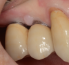
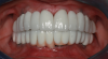
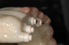
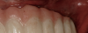
There are a wide variety of such interdental products on the market. For implant patients, the author recommends choosing an oral irrigator that pulsates and has at least two tips: the universal tip and the dental implant tip with three tufts. A Tufts University School of Dental Medicine study found a water flosser with implant tip was 145% more effective than string floss around implants when used in conjunction with a manual toothbrush.9 Significant reduction of gingival bleeding at 2 weeks and 30 days, respectively, were reported.9 Improvement in the health of tissues around implants can be seen in one patient who presented with inflammation around an implant (Figure 1), and another patient with a full-arch prosthesis that now has minimal-to-no plaque around both the top and bottom peri-implant tissues (Figure 2).
An oral health study from the University of Southern California’s Center for Biofilm Research reported that holding a water jet over the surface of the implant tooth removed 99.9% of mature plaque biofilm in only 3 seconds.10
Establishing a Good Implant Maintenance Program
Brushing and Flossing
Recommendations to patients should consider the individual—including dexterity, motivation, and the type of restoration. Unless there are dexterity problems, the author generally leaves it to implant patients to choose whether to use an electric or manual toothbrush, but based on the literature, strongly recommends that they incorporate a water flosser or oral irrigator. Patients who are avid string flossers should be cautioned about the risk of injury to gingiva and peri-implant tissues using the floss because the attachment of the peri-implant tissues is not as strong as the periodontium.11,12 To prevent permanent damage to those tissues, they may be switched to an oral irrigator.
Educating Patients About the Importance of Maintenance
In addition to the challenge of convincing patients to adhere to proper home maintenance is getting them to comply with recommended office visit recommendations. For that reason the team must take the time to educate patients about not only the methods, but the importance of the maintenance visit. Patients should be made aware that this is not just to prevent peri-implantitis—which can be costly, time-consuming, and unpredictable—but to maintain their oral health for the sake of their overall health, especially if they are suffering from or are at risk for chronic illnesses.
Recall Visit Management for Implant Patients
Intervals Between Appointments
Determining the interval between appointments—anywhere from 1 to 6 months—should be personalized, based on the patient’s ability to perform good oral hygiene at home. There is no protocol established in the literature for the 6-month interval for implant patients. To prevent the development of peri-implantitis in a patient who has difficulty cleaning underneath the prosthesis, an interval of 1 or 2 months may be appropriate.
Length of Appointments
Then, too, to consider is the length of the appointment—anywhere from 30 to 60 minutes, depending on the complexity of the care rendered in the office. For example, for a patient with six implants and no dentition, 30 minutes may be sufficient, but most patients have mixed dentition and implants, so usually a 45- to 60-minute dental maintenance visit is necessary in order to complete good record-taking, patient education, and plaque removal.
Implant-Related Record-Keeping
Records such as photographs and radiographs taken by the hygienist at the implant maintenance appointment show how the patient is maintaining over time. Ideally, radiographs should be taken once a year. Pocket depths, BOP, inflammation, and plaque levels should be reported. Occlusion and mobility also should be noted. Baseline photographs, radiographs, and probing depths should be taken at the time of implant placement and prosthesis delivery, so changes can be monitored over time.
Medical History, Including Supplements
In addition to assessing the patient’s hygiene, oral health, systemic health, and medications, it is important to determine their wellness status and ask about nutritional supplements, because 68% of Americans today take some kind of supplements that can affect oral health.13 If they do not take supplements, that could be a problem, too. An example is a lack of vitamin D—a systemic deficiency that increases the risk of osteoporosis, high blood pressure, allergies, colds and flu, mental health, and heart problems. An adequate level, on the other hand, positively affects oral health by reducing inflammation and modulating cell growth and immune function. Vitamin D level has also been inversely associated with gingival bleeding and level of periodontal disease.14
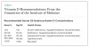
Therefore, the author suggests gathering information about the patient’s vitamin D level or asking them to be tested. Optimal vitamin D recommendations (Table 1) are now rising in keeping with research that supports it.14
Radiographs
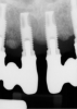
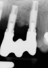
Given patient concerns about radiation exposure, it is important to know how, when, and why radiographs should be taken. Although there are concerns about radiation, radiographs make it possible to determine bone levels at baseline—ie, at implant and restoration insertion—and to monitor them over time. Vertical radiographs should be as parallel as possible to the implant body, so that the implant threads are clearly visible. Typically, periapicals should be taken once a year around dental implants. Figure 3 and Figure 4 shows bone loss up to the fifth and sixth thread.
Probing
It is important to know when and how to probe for the same reasons as taking radiographs—to establish initial pocket depths at baseline (insertion of the restoration) and to monitor for changes over time. For this, a traditional periodontal probe should be used, exerting light force, 0.25 N. However, unlike bone loss, neither BOP nor probing depths—even as much as 6 mm—are correlated to bone loss around implants, according to the Estepona consensus on peri-implantitis 2012.15
Assessing Plaque Level
The plaque/calculus level should also be documented regularly for signs of changes. The location (supragingival, subgingival, or on the prosthesis itself), severity (mild, moderate, severe), and whether it is anterior/posterior, buccal/lingual, and upper should be documented to see if there is improvement at the next hygiene visit.
According to the literature, the location of plaque may affect its accumulation. Quirynen et al16 found minor differences in the amount of plaque on rough versus smooth surfaces, but Wennerberg et al17 reported similar plaque formation. Baldi et al concluded that dual acid-etched surfaces have greater plaque accumulation than machined surfaces, but there is less marginal bone loss.18 Because etched or rough surfaces around dental implants have greater plaque accumulation but less marginal bone loss than machined surfaces, it is best to try to protect implant surfaces from plaque exposure but to correct it if it has been exposed.
Plaque Accumulation Removal
While some hygienists like to use plastic scalers, studies show that all scalers, whether filled or unfilled resin or titanium, leave scratches on the titanium surface.19 Instead, hygienists can use ultrasonic devices that come with the rubber tip protectors that cause less damage or scratches to the abutment. Hygienists should be mindful of minimizing scratches on the smooth implant/abutment surface, because scratches can actually increase the plaque accumulation.
The author does not recommend using air-powder abrasive for cleaning around implants to avoid bleeding particles around the tissues. A study by Tastepe and others noted minor surface changes on titanium treated with air-powder abrasives, with treatment results influenced by the powder type being used, the application time, and whether the powder was applied surgically or nonsurgically; remnants were observed on and impacted in the titanium surface.20
Checking Occlusion/Occlusal Overload
That the implant patient’s occlusion should be checked at every hygiene visit is suggested by the literature, including a monkey study by Isidor associating a loss of osseointegration with occlusal overload of oral implants21 and a review by Fu et al22 in which unbalanced occlusion affected the marginal bone loss around dental implants. A study by Miyata et al,23 which examined the influence of experimental occlusal overload on peri-implantitis in monkeys, showed that bone resorption around implants tended to increase with 180 µm or more excessive height of the superstructure, suggesting that minor occlusal overload is not a significant concern. However, over time—especially with mixed dentition, eg, porcelain or zirconia crowns, which wear differently than enamel—the implant can suddenly be in hyperocclusion associated with bone loss, which could eventually lead to implant loss. Therefore, occlusal overload should be checked and, if necessary, adjusted every 6 months to prevent problems.
Dealing With Residual Cement
Residual cement left subgingivally can lead to bone loss and implant failure, as observed by Wilson,24 whose findings support scrupulous cement removal at the time of cementation, considering the unintended presence of cement as a potential cause when signs of peri-implant disease are seen. Ideally, whenever possible, a screw-retained restoration should be fabricated. If cementation is unavoidable due to the angle of the implant, then it is important to use a radio-opaque cement, which is visible on x-rays, to facilitate complete removal.25 It is also important that the margins of the implant crown are not placed subgingivally to allow for visualization of the cement and easy removal.
Bacterial Culture
The literature offers some insight about whether or not to culture patients who have peri-implantitis or some slight suppuration from mucositis. A study by Renvert et al26 did not find any significant difference between the bacteria in nearly 1,000 implants, whether or not affected by peri-implantitis. These findings suggest peri-implant disease is a factor of the amount of bacteria and the genetic susceptibility of the patient, not necessarily the type of bacteria that is in the mouth. Similarly, Hultin et al27 found high levels of periodontal pathogens in peri-implantitis.
Both studies suggest that it is not necessary to culture right away. Most important is, if there is a significant amount of suppuration inflammation, the area should be irrigated locally with antimicrobials; systemic antibiotics are really not necessary. In short, culturing should be done on a case-by-case basis, not for everyone with peri-implantitis.
Understanding Prosthesis Contours and Cleansability
The contours of the prosthesis can have a significant impact on the patient’s ability to maintain good oral hygiene at home and keeping healthy peri-implant tissues. The practitioner should be aware of this issue and make sure restorations are as cleansable as possible. For example, a concave surface from the abutment to the buccal surface is a plaque trap and should be immediately corrected (Figure 5 and Figure 6).
It is particularly important to ensure that the space beneath the prosthesis be designed to maximize cleansability. To prevent food impaction, a gap beneath the prosthesis should be minimized through correction, such as realignment. In general, all intaglio surfaces of the prosthesis should be convex or flat, and buccolingual dimensions should be as narrow as possible, but not so thin that the prosthesis is likely to break. The patient must be able to get the water flosser all the way through and underneath the prosthesis.
The clinician should also be aware of the importance of having keratinized attached tissue around dental implants, and monitor for it. Thicker keratinized attached gingiva creates a better seal around the abutment and prevents plaque and food accumulation.
Using Ozone
To combat fungus, bacteria, and parasites that may contribute to peri-implant disease, the author has incorporated ozone in recent years. This ozone, which is three molecules of oxygen that are used in the form of gas or ozonated water to clean in both dentistry and medicine, safely helps kill any fungus, bacteria, or parasite on contact. However, although it is natural and safer than bleach, because the only two tissues in the human body that do not have anti-oxygen capacity are the eyes and the lungs, it should not be inhaled, and eye protection is needed. Proper training is required if ozone is to be incorporated in the dental office.
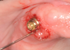
Ozone is used in peri-implantitis or where the beginning of inflammation is seen in mucositis by using a cannula (Figure 7) to blow the gas directly underneath the tissues affected. Although this has been around for more than 100 years, until recently there were few studies, likely due to the advent of antibiotic resistance to antimicrobials. However, one that studied the influence of gaseous ozone in peri-implantitis reported that a 24-second treatment around areas of the inflammation can sterilize and eliminate the microorganisms Strep sanguinis and Porphyromonas gingivalis without affecting the osteoblasts and the tissue cells.28
Improving Hygiene Compliance
There are several components to improving patients’ hygiene compliance: understanding their desires; identifying causes of noncompliance; making hygiene visits pleasurable; and changing their protocol and getting them involved in decisions made. These concepts apply to all patients, but the stakes—uncontrolled disease leading to implant failure—are especially high for implant patients. Therefore, the dental team needs to help them understand why it is important to recognize initial mucositis or initial changes because it is more predictable to correct them in the beginning. It is important to work with patients who are not receptive to that message or are negligent about their hygiene appointments. A protocol designed in keeping with their desires and abilities and that is developed with their involvement is most likely to be effective. For example, a patient who cannot or will not use string floss should be changed to an oral irrigator, and those unwilling or unable to practice optimal homecare should be seen in the office more frequently.
Conclusion
Optimal peri-implant maintenance is based on several pillars. First, the clinicians and all dental team members must listen carefully to each patient to understand their desires and goals. Based on individual patients’ abilities and needs, implant patients should be educated on a personalized program of home care. Further, the dental team should ensure that hygiene interval schedules and appointment lengths are customized to meet the individual patient’s needs.
About the Author
Dr. Moldovan is a periodontist and nutritionist whose practices in Los Angeles, California, and New York City are primarily devoted to implant therapy.
Disclosure
Dr. Moldovan received an honorarium for the preparation of this manuscript.
References
1. Froum SJ, Rosen PS. A proposed classification for peri-implantitis. Int J Periodontics Restorative Dent. 2012;32(5):533-540.
2. Atieh MA, Alsabeeha NH, Faggion CM Jr, Duncan WJ. The frequency of peri-implant diseases: A systematic review and meta-analysis. J Periodontol. 2013;84(11):1586-1598.
3. Daubert DM, Weinstein BF, Bordin S, et al. Prevalence and predictive factors for peri-implant diseases and implant failure: a cross-sectional analysis. J Periodontol. 2015;86(3):337-347.
4. Truhlar RS, Morris HF, Ochi S. The efficacy of a counter-rotational powered toothbrush in the maintenance of endosseous dental implants. J Am Dent Assoc. 2000;131(1):101-107.
5. Louropoulou A, Slot DE, Van der Weijden F. Mechanical self-performed oral hygiene of implant supported restorations: a systematic review. J Evid Based Dent Pract. 2014(Jun);14 Suppl:60-69.e1.
6. Grusovin MG, Coulthard P, Worthington HV, Esposito M. Maintaining and recovering soft tissue health around dental implants: a Cochrane systematic review of randomised controlled clinical trials. Eur J Oral Implantol. 2008;1(1):11-22.
7. Magnuson B, Harsono M, Stark PC, et al. Comparison of the effect of two interdental cleaning devices around implants on the reduction of bleeding: a 30-day randomized clinical trial. Compend Contin Educ Dent. 2013;34(Spec No 8):2-7.
8. Ioannidis A, Thurnheer T, Hofer D, et al. Mechanical and hydrodynamic homecare devices to clean rough implant surfaces - an in vitro polyspecies biofilm study. Clin Oral Implants Res. 2015;26(5):523-528.
9. The Effect of a Water Flosser with Plaque Seeker Tip® on Gingival Bleeding for Implant Patients. Presented at the International Association of Dental Research, Seattle, WA, USA. March 23, 2013. Abstract #3761.
10. Gorur A, Lyle DM, Schaudinn C, Costerton JW. Biofilm removal with a dental water jet. Compend Contin Educ Dent. 2009;30(Spec No 1):1-6.
11. Misch, CE. Contemporary Implant Dentistry. 3rd ed. St. Louis, MO: Mosby Elsevier; 2008:75-76.
12. Hallmon WW, Waldrop TC, Houston GD, Hawkins BF. Flossing clefts. Clinical and histologic observations. J Periodontol. 1986;57(8):501-504.
13. Council for Responsible Nutrition press release. Most Americans take—and trust—supplements. New Hope Network website. October 23, 2015. http://newhope.com/news-analysis/most-americans-take-and-trust-supplements. Accessed April 25, 2016.
14. Moldovan S. Update on nutrition for better healing. Inside Dentistry. 2015;11(7):45-52.
15. Albrektsson T, Buser D, Chen ST, et al. Statements from the Estepona consensus meeting on peri-implantitis, February 2-4, 2012. Clin Implant Dent Relat Res. 2012;14(6):781-782.
16. Quirynen M, van der Mei HC, Bollen CM, et al. An in vivo study of the influence of the surface roughness of implants on the microbiology of supra- and subgingival plaque. J Dent Res. 1993;72(9):1304-1309.
17. Wennerger A, Sennerby L, Kultje C, Lekholm U. Some soft tissue characteristics at implant abutments with different surface topography. A study in humans. J Clin Periodontol. 2003;30(1):88-94.
18. Baldi D, Menini M, Pera F, et al. Plaque accumulation on exposed titanium surfaces and peri-implant tissue behavior. A preliminary 1 year clinical study. Int J Prosthodont. 2009;22:447-455.
19. Hasturk H, Nguyen DH, Sherzai H, et al. Comparison of the impact of scaler material composition on polished titanium implant abutment surfaces. J Dent Hyg. 2013;87(4):200-211.
20. Tastepe CS, van Waas R, Liu Y, Wismeijer D. Air powder abrasive treatment as an implant surface cleaning method: a literature review. Int J Oral Maxillofac Implants. 2012;27(6):1461-1473.
21. Isidor F. Loss of osseointegration caused by occlusal load of oral implants. A clinical and radiographic study in monkeys. Clin Oral Implants Res. 1996;7(2):143-152.
22. Fu JH, Hsu YT, Wang HL. Identifying occlusal overload and how to deal with it to avoid marginal bone loss around implants. Eur J Oral Implantol. 2012;5(Suppl):S91-S103.
23. Miyata T, Kobayashi Y, Araki H, et al. The influence of controlled occlusal overload on peri-implant tissue. Part 3: A histologic study in monkeys. Int J Oral Maxillofac Implants. 2000;15(3):425-431.
24. Wilson TG Jr. The positive relationship between excess cement and peri-implant disease: A prospective clinical endoscopic study. J Periodontal. 2009;80(9):1388-1392.
25. Burbano M, Wilson TG Jr, Valderrama P, et al. Characterization of Cement Particles Found in Peri-implantitis-Affected Human Biopsy Specimens. Int J Oral Maxillofac Implants. 2015;30(5):1168-1173.
26. Renvert S, Roos-Jansåker AM, Lindahl C, et al. Infection at titanium implants with or without a clinical diagnosis of inflammation. Clin Oral Implants Res. 2007;18(4):509-516.
27. Hultin M, Gustafsson A, Hallström H, et al. Microbiological findings and host response in patients with peri-implantitis. Clin Oral Implants Res. 2002;13(4):349-358.
28. Hauser-Gerspach I, Vadaszan J, Deronjic I, Gass C, et al. Influence of gaseous ozone in peri-implantitis: bactericidal efficacy and cellular response. An in vitro study using titanium and zirconia. Clin Oral Investig. 2012;16(4):1049-1059.